Use of Electron Microscopy (EM)to Detect Transplant Glomerulopathy (TG) in Patients with Late-Indication Allograft Biopsy
Rafael Maldonado1, Maria Fernanda Toniolo2, Matias Bina1, Marisol Rodriguez1, Santiago Carbel1, Carolina Zaguirre1, Natalia Matamala1, Maria Florencia Rodriguez1, Paula Dionisi1, Carlos Idoria1.
1Nephrology and Transplantation Service, Clinica Privada Velez Sarsfield, Cordoba, Argentina; 2Renal Pathology Service, Centro Diagnostico Patologico SRL, Buenos Aires, Argentina
Introduction: The main causes of late graft loss are: death of patients with functioning graft and chronic allograft nephropathy, in which it stands out, to chronic rejection mediated by antibodies (cABMR). cABMR was defined during the Banff meeting in 2005 due to: Transplant Glomerulopathy (TG), Multilamination of the basal membrane of the tubular pericapillary (MLMBPCT), Transplant vasculopathy and interstitial fibrosis with tubular atrophy (IFTA), with deposit of C4d in PCT and the presence of Anti-HLA Antibodies specific donor (DSA).
The presence of C4d deposition in the peritubular capillary (PTC) is a diagnostic marker of ABMR greater than 50% in acute graft rejection but not in its chronic or late ABMR phenotype, so its usefulness is currently questioned in the late phase, identifying them as C4d negative.
The typical characterizes cABMR is TG recognized as a glomerulopathy that manifests a double contour of the glomerular capillary,. The cABMR with duplication or MLMBG, increase of the subendothelial space, interposition of mesangial cells, loss of fenestration in the endothelial cells, and MLMBPTC as the findings most significant effects linked to the interaction of antibodies in the glomerular endothelium.
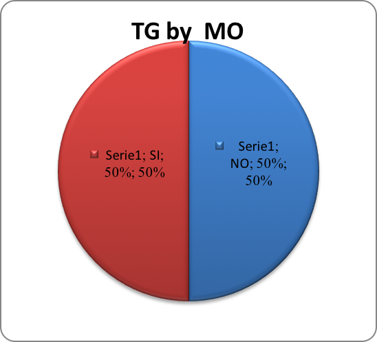
Objectives: Detect morphological changes of TG by ME in patients with chronic allograft dysfunction who have late biopsies for clinical indication, whose previous histopathological evaluation was by Banff classification in MO and IHC for C4d.
Materials and Methods: We performed a retrospective observational study of a descriptive cohort, kidney transplant patients performed in the Nephrology and Transplant Service of the CPVS, Córdoba, Argentina who presented chronic allograft dysfunction with late biopsies for clinical indication performed in the period from July 2011 to August 2016.The MO and ME analysis was performed by two different pathologists.The indication for renal biopsy was performed according to the following criteria: increase in serum creatinine ≥0.3 mg / dl or more during the post-transplant evolution. Proteinuria evaluated by Pr/Cr ratio in spot urine ≥0.3 mg / dl.3. Patients with at least 6 months of kidney transplant.4. All samples of biopsies performed should have at least 4 glomeruli for the MO and ME analysis, respectively.
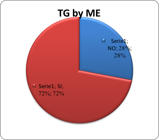
Results: Inflammation of the glomerular endothelium (GECS) in 21/22 95%.Podocyte deletion processes in 100% of patients, 22.7% severe and in the rest 76% mild to moderate. MLMBPTC, 68% were present in 63.6% 14/22 patients mild (<3 layers) and in only 5% was moderate (7 layers). ). MLMBG in 17/22 77.2%. 

Conclusion: The use of EM in late kidney transplant biopsies by indication allowed detecting ultrastructural changes of transplant glomerulopathy of cABMR in a high % of patients with C4d negative w/wo DSA. Prospective studies should be necessary as protocol biopsies with earlier changes suggestive of TG with EM, and anticipate the clinically evident damage.
